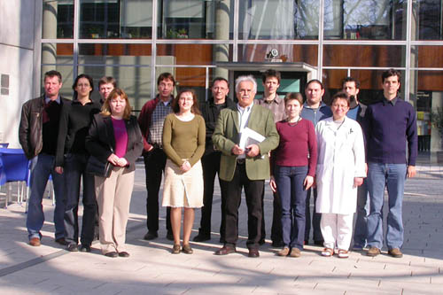
From left to right: Zahuczky, Gábor; Vecsei Zsófia; Király, Róbert; Darai, Zoltánné; Mádi, András; Hodrea, Judit; Balajthy, Zoltán; Prof. Fésüs, László, Csomós, Krisztián; Csősz, Éva; Németh, István; Klem, Attiláné; Doró, Zoltán; Keresztes, Gábor
Ever dreamed about a car that would not only maintain but also repair itself without the need to
In human the concerted activity of more than 10^15 cells will make sure that the body functions as intended. Such a complex structure can only function if individual cell fate is subdued in favor of the general interest of the organism. The principal mechanism ensuring that the interest of organism prevails over the interest of a cell is that the cell cycle is tightly regulated. Most cells can only thrive if they receive signals to do so. In the absence of these trophic signals cells undergo various forms of natural cell death also called apoptosis.
Natural cell death is accompanied with changes on the cell surface that allow the recognition and clearance of these cells. Apoptotic cells are removed from the tissues by engulfment in a process called phagocytosis. Uptake of apoptotic cells, recently referred to by some as efferocytosis, in most cases is likely to be performed by adjacent non-apoptotic cells, "non-professional phagocytes" as well as resident "professional phagocytes" (e.g. dendritic cells). If the capacity of these cells to take up apoptotic cells is exceeded, a frequent scenario during development and regeneration of tissues after infection or injury, macrophages, a class of "professional phagocytes" are recruited. The clearance is usually so rapid and efficient that apoptotic cells cannot be easily detected in most tissues.
The situation is complicated by the presence of extrinsic factors. Pathogens will try to take advantage of the resources and constant milieu of the host organism, and may directly or indirectly cause the affected host cells to perish. Noxious environmental stimuli (excess heat, mechanical wear, toxic substances) may lead to the same outcome. Inorganic particles may also get to or form within the tissues, which might disrupt the normal function of tissues and lead various forms of "unnatural" cell death, necrosis, anoikis or autophagic cell death.
Professional phagocyte populations are heterogeneous in terms of their ability to phagocytose various classes of particulate material. The molecular mechanisms underlying this phenomenon are not completely understood. By developing methods to separate macrophages that are and are not capable of phagocytosing various classes of particulate material and comparing them with genomic and postgenomic tools we aim to get a better insight of the molecular mechanisms governing phagocytosis.
The clearance process must not only be effective, but also provide the cell types responsible for the clearance with signals on the state of the tissue. These signals will integrate to anywhere between a full-fledged anti-inflammatory response to a vigorous inflammatory response at the organism level, depending on whether particles to be cleared are predominantly apoptotic cells or fall into other categories.
There are modifiers of the stereotypic inflammatory and anti-inflammatory response at the organism level. Several nuclear hormone receptors that are primarily involved in regulating metabolism have been implicated in the organism-level orchestration of inflammatory and anti-inflammatory responses. For example glucocorticoids acting through the glucocorticoid receptor, a member of the nuclear hormone receptor family have long been recognized for their potent anti-inflammatory activities. Glucocorticoids and glucocorticoid analogs acting through nuclear hormone receptors are known to be potent anti-inflammatory drugs. Glucocorticoid treatment during early steps of the macrophage differentiation was shown to enhance the ability of macrophages to engulf apoptotic cells. This suggests that the ability of macrophages to efferocytose is transcriptionally regulated. We are interested in deciphering the transcriptional changes that lead to the increased efferocytosis upon glucocorticoid treatment.
Szondy, Zs., Sarang, Zs., Molnár, P., Németh, T., Piacentini, M., Mastroberardio, P.G., Falasca, L., Aeschlimann, D., Kovács, J., Kiss, I., Szegezdi, E., Lakos, G., Rajnavölgyi, É., Birckbichler, P.J., Melino, G. and Fésus, L. (2003): TGASE2-/- mice reveal a phagocytosis-associated crosstalk between macrophages and apoptotic cells. Proc. Natl. Acad. Sci. USA 100, 7812-7817
Gy. Májai, G. Petrovski and L. Fésüs (2005): Inflammation and the apopto-phagocytic system. Immunology Letters 104, 94-101
Tissue transglutaminase has long been implicated to play a pivotal role in apoptosis. Using TG2 knockout mice we demonstrated that the lack of TG2 leads to impaired ability of mouse macrophages to phagocytose apoptotic cells. The exact mechanism by which TG2 exerts its activity is currently under investigation in collaboration with the Apoptosis Signaling group of the institute.
Interestingly the lack of TG2 in neutrophils leads to increased phagocytosis of pathogens. Pathogens and apoptotic cells fall into different phagocytosis classes, and the roles of mononuclear and polymorphonuclear phagocytes are very different. The lack of TG2 also leads to abnormal oxidative response and defective migration kinetics.
As an independent model to study the role of this enzyme we use the all-trans retinoic acid (ATRA) induced cell differentiation process of the promyelocytic NB4 leukemia cells. ATRA drives the NB4 cells to differentiate into neutrophil-granulocyte-like cells. In this process TG2 gets up-regulated both at the transcriptional and translational levels. Furthermore we provided evidence that part of the TG2 translocates into the nucleus of differentiating NB4 cells. Inhibiting TG2 with transglutaminase inhibitors or knocking down the TG2 expression by siRNA will result in an altered differentiation process.
The exact molecular mechanisms by which TG2 can contribute to or mediate functions of neutrophil or macrophage are presently unknown. Our results show that TG2 may both modulate cell signaling and alter gene expression patterns.
Recent selected publications:
L. Fésüs, Z. Szondy (2005): Transglutaminase 2 in the balance of cell death and survival. FEBS Letters 579, 3297-3302
Balajthy Z., Szantó A., Vámosi Gy., Csomós K., Lanotte M., Fésüs L. (2006) Tissue-transglutaminase contributes to neutrophil granulocyte differentiation and functions. Blood, 2006; 108: 2045-2054
Keresztessy, Z, Csosz, E, Harsfalvi, J, Csomos, K, Gray, J, Lightowlers, RN et al., (2006) Phage display selection of efficient glutamine-donor substrate peptides for transglutaminase 2. Protein Sci 15: 2466-80
Celiac disease
issue transglutaminase has been shown to the major autoantigen in coeliac disease.
Collaborators: Korponay-Szabó Irma
Recent selected publications:
I.R. Korponay-Szabó, K. Laurila, Z. Szondy, T. Halttunen, Z. Szalai, I. Dahlbom, I. Rantala, J.B. Kovács, L. Fésüs, M. Maki: Missing endomysial and reticulin binding of coeliac antibodies in transglutaminase 2 knockout tissues. Gut 52, 199-204 (2003)
I.R. Korponay-Szabó, T. Halttunen, Z. Szalai, K. Laurila, R. Király, J.B. Kovács, L. Fésüs, M. Maki (2004): In vivo targeting of intestinal and extraintestinal transglutaminase 2 by coeliac autoantibodies. Gut 53, 641-648

